Emergency Thoracotomy
(From BMJ Journals, 2005, Wise et al. )
Background
– Indication is penetrating chest/epigastric trauma associated with cardiac arrest (any rhythm)
– Should be performed within 10 minutes of loss of cardiac output
– Contraindications:
o Definitive loss of cardiac output more than 10 minutes
o Any patient with cardiac output, including hypotensive patients
o Blunt trauma
– Total time should be 2-3 minutes
Equipment
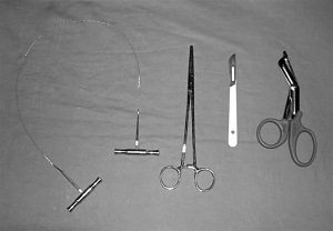
Gigli saw
Large clamp/forceps
Large scalpel
Large scissors
Procedure:
1. Position in supine position
2. Intubation, ventilation and iv acess should be performed synchronously, don’t wait for them to happen before starting!
3. Rapid (if any) prepping of skin
4. Use scalpel and blunt forceps to make 4cm thoracostomies in both sides of the chest (breaching intercostal muscles and parietal pleura) in the 5th intercostal space in mid-axillary line (as for chest drains)
a. If a tension pneumothorax is released and cardiac output returns, stop here
5. Insert 2 fingers into the thoracostomy to hold lung out of the way and cut through all layers to the sternum with heavy scissors
6. Cut through sternum or xiphoid with the heavy scissors, and if not able to, use gigli saw.
a. Pass forceps under the sternum, grasp one end of the giggle saw and pull through to the other side
b. Connect saw handles
c. Use smooth long strokes to cut through the bone
7. Open the “clam shell” using rib-spreaders and if not available, gloved assistants. Open to the maximum exposure
8. Lift the pericardim with forceps and make a large midline longitudinal incision with scissors (minimises damage to phrenic nerves, which run along lateral walls of pericardial sac)
9. Evacuate all blood and clot then inspect the heart rapidly and systematically for site of bleeding
a. If cardiac output returns spontaneously, close cardiac wounds
b. If heart beat is slow with significantly reduced CO, close wounds quickly then supplement with internal cardiac massaging and inotropic support
c. If heart remain in asystole, close wounds quickly and attempt internal cardiac massage. Just “flicking” the heart can return contractions.
Internal cardiac massaging:
– Can be single handed, or preferably, place one hand behind posterior surface of the heart and one on the anterior surface.
– “Milk” blood upwards from the apex at a rate of 80/min
– Ensure the heart remains horizontal during massaging (if lifted, reduces venous filling)
– An assistant can compress the aorta against the spinal column using a thumb or fingers to maximise coronary and cerebral perfusion
Controlling bleeding
– Holes less than 1cm can be occluded temporarily with finger or gauze swab. If successful, don’t attempt other methods
– For larger defects, a Foley urinary catheter can be inflated through the hole and pulled back. Only use a small (10ml) volume in balloon to allow sufficient stroke volume. Ensure catheter is clamped to stop blood exiting through it.
o If used, a giving set can be connected through the catheter to give blood/fluid directly into the heart
– IF bleeding cannot be controlled as above, close with large sutures AS A LAST RESORT (risk of occluding coronary arteries). Use the minimum number necessary to allow finger/gauze/Foley to close defect.
o Non-absorbable 0/0 or 1/0
o Monofilament or braided
o Take 1-2cm bites
If defibrillation necessary, use internal pads with initial level of 10 joules.
– If not available, close the clamshell and use external pads
If procedure is successful,
– bleeding will begin from internal mammary and intercostal vessels. Control large bleeders with artery forceps.
– Give sufficient analgesia as patient might wake up
– Move patient to theatre (ideally in cardiothoracic facility) for definitive repair.